Skip to content

1970/01

HIV/AIDS

Source: Pocket Medicine, 2022

up:: 專師考試解題

定義與臨床表現

  • 急性HIV:皮疹、淋巴結腫大、發燒、口腔潰瘍、咽喉炎、肌肉疼痛、腹瀉
    • 一般在接觸後約2-6週出現;並非所有HIV感染者會出現急性HIV的症狀
  • 愛滋病(AIDS):HIV + CD4 <200/mm³ 或出現AIDS定義的伺機性感染(OI)或惡性腫瘤

HIV 感染中定義愛滋病(AIDS)的伺機性感染與指標性疾病

  • HIV 引起的腦病變(Encephalopathy attributed to HIV)
  • 進行性多灶性白質腦病變(Progressive multifocal leukoencephalopathy)
  • 多次或反覆性細菌感染(僅限 6 歲以下兒童)
  • 沙門氏菌敗血症,反覆發作
  • 肺炎,反覆發作(僅限成年人、青少年及年滿 6 歲以上兒童)
  • 支氣管、氣管或肺部念珠菌感染
  • 食道念珠菌感染
  • 播散性或肺外球孢子菌病(Coccidioidomycosis)
  • 肺外隱球菌病(Cryptococcosis)
  • 播散性或肺外組織胞漿菌病(Histoplasmosis)
  • Pneumocystis jirovecii(舊稱 P. carinii)肺炎
  • 巨細胞病毒感染(不含肝、脾或淋巴結),發病年齡 >1 個月
  • 巨細胞病毒視網膜炎(合併視力喪失)
  • 單純皰疹病毒感染:慢性潰瘍(持續 >1 個月)或支氣管炎、肺炎、食道炎(發病年齡 >1 個月)
  • 播散性或肺外 Mycobacterium avium complexMycobacterium kansasii 感染
  • 任一部位之結核病(Mycobacterium tuberculosis):肺結核(僅限成年人、青少年與年滿 6 歲以上兒童)、播散性或肺外結核
  • 播散性或肺外感染的其他分枝桿菌或未鑑定分枝桿菌
  • 侵犯性子宮頸癌(僅限成年人、青少年與年滿 6 歲以上兒童)
  • 卡波西肉瘤(Kaposi sarcoma)
  • 伯基特淋巴瘤(Burkitt lymphoma 或同等類型)
  • 免疫母細胞淋巴瘤(Immunoblastic lymphoma 或同等類型)
  • 原發性中樞神經系統淋巴瘤 (Primary CNS lymphoma)
  • HIV 引起的惡病質症候群(Wasting syndrome attributed to HIV)
  • 慢性腸道隱孢子蟲病(Cryptosporidiosis)(持續 >1 個月)
  • 慢性腸道等孢子蟲病(Isosporiasis)(持續 >1 個月)
  • 腦部弓形蟲病(Toxoplasmosis of brain),發病年齡 >1 個月

流行病學

  • 約有120萬美國人與HIV感染共存(13% 不知道自己感染);全球約3700萬
  • 高風險群體:男性同性戀者、變性女性、靜脈注射毒品者、性工作者、高風險患者的伴侶
  • 傳播途徑:性行為(無抗病毒藥物時風險為0.1-1%)、針扎(職業或靜脈注射毒品)、垂直傳播(無抗病毒藥物時風險為15-40%)、輸血、器官移植(在美國不常見)

預防 (NEJM 2015;373:2237; Lancet 2016;387:53; J Infect Dis 2018;218:16; CDC 2021)

  • 暴露前預防(PrEP):每日服用TDF/FTC,若遵守療程可減少超過90%的傳播風險。考慮用於HIV陰性伴侶、無安全套性行為的高風險群體、6個月內有性傳播感染史、靜脈注射毒品且共用針頭者。開始之前需排除HIV,並每3個月檢查腎功能、性病及HIV。
  • 暴露後預防(PEP):在高風險暴露後盡快(72小時內)開始(如果HIV狀態不明則需要個別決定)。檢查基礎HIV、性病、HBV、HCV。治療:2種NRTI(通常是TDF/FTC)+ RAL或DTG × 4週。考慮後續開始PrEP。

篩檢與診斷 (JAMA 2018;320:379)

  • 所有13-64歲人群至少篩檢一次,每次懷孕時、若有新性病感染診斷時篩檢;高風險者每年篩檢
  • HIV抗體/p24抗原(ELISA檢測):感染後1-12週陽性;靈敏度超過99%;為主要篩檢測試
  • 如果結果為陽性,抗體分型檢測可確認並區分HIV-1與HIV-2(MMWR 2013;62:489)
  • 血漿HIV RNA PCR病毒量(VL):檢測範圍為20-1000萬拷貝/mL;偽陽性可能發生,但通常拷貝數量較少;相比之下,初期感染病毒量應該較高(>75萬)
  • CD4計數:不是診斷測試,因為可能HIV陽性但CD4正常,或HIV陰性但CD4低

對新診斷HIV陽性患者的處理 (CID 2020;73:e3572)

  • 諮詢關於若服藥遵從性高則治療預後良好、治療選擇及病情披露
  • 實驗室評估:CD4計數、HIV病毒量及基因型、CBC/DC、基礎生化、肝功能、HbA1c、血脂肪、尿液分析、PPD/IGRA、梅毒抗體、披衣菌與淋病(3部位)、A/B/C型肝炎、G6PD(如果是高風險族群)、懷孕篩檢、若使用abacavir則檢查HLA-B*5701
    • 如果是AIDS:CMV IgG、Toxo IgG。
  • 確認所有疫苗(包括每年流感疫苗)都是最新的,若CD4 ≤200則避免使用活菌疫苗
  • 及早開始抗病毒藥物(最好在實驗室檢查和基因型結果後,同時得到HIV專家指導),不論CD4數值如何,因為可以降低死亡率(NEJM 2015;373:795)
  • 治療可以預防對伴侶的傳播。當病毒量檢測不到超過6個月時,無保護性行為的傳播風險約為0%(JAMA 2016;316:171;Lancet HIV 2018;5:e438)。
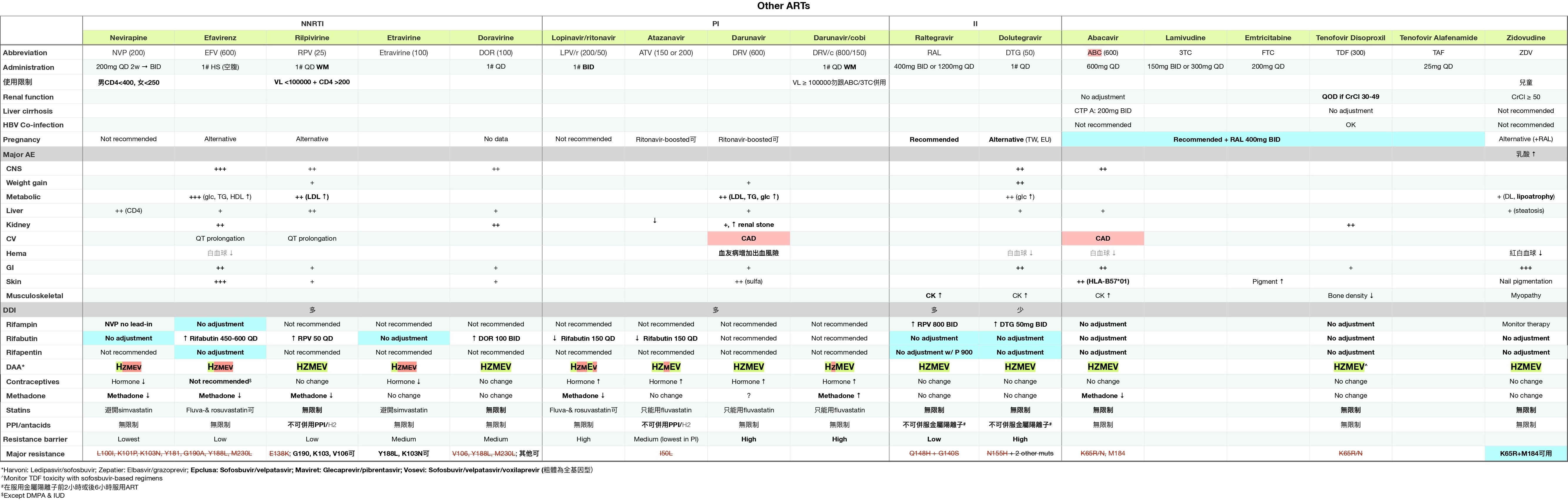
  • 治療方案包括:2種NRTI(如TAF + FTC)+ INSTI或加強型PI(如DRV/r)
  • 開始抗病毒治療可能會因免疫重建發炎症候群(IRIS)暫時加重現有的伺機性感染(TB、MAC、CMV等)。在抗病毒治療的前4週使用類固醇可降低TB相關IRIS的風險,但不建議常規使用(NEJM 2018;379:1915)。
  • 如果擔心有隱球菌或TB腦膜炎,不應立即開始抗病毒治療
  • 開始抗病毒治療後,需每4週檢查一次病毒直到達到不可檢測,之後每3-4個月監測一次

對之前已知HIV陽性患者的處理

  • 病史與體檢(黏膜皮膚、神經認知、伺機性感染、惡性腫瘤、性病);藥物及依從性
  • 回顧抗病毒藥物(過去和現在);如果住院,通常繼續使用抗病毒藥物,如果必須停用,則全部停止以降低抗藥性風險
  • 治療失敗:在使用抗病毒藥物幾個月後無法達到不可檢測VL、病毒反彈(在之前的抑制後VL >200拷貝/mL ×2次)、CD4計數下降或臨床惡化

機會性感染預防

感染 指徵 初級預防
結核 PPD陽性(≥5 mm)、IGRA或高風險暴露 參見潛伏結核的治療
肺囊蟲肺炎_Pneumocystis jiroveci_ (PCP) CD4 <200/mm 或 CD4 <14%口腔念珠菌感染 TMP-SMX(首選)或dapsone或atovaquone或吸入pentamidine
隱球菌 CD4 <150/mm 且有地方流行/暴露史 Itraconazole
弓形蟲 CD4 <100/mm³ 且Toxo IgG陽性 TMP-SMX或dapsone 50 mg + pyrimethamine + leucovorin
MAC 若已開始有效抗病毒藥物則不再建議預防
停止預防的時間:若CD4 >200 × 3個月則停止PCP和弓形蟲的預防

HIV/AIDS的併發症

CD4 計數 併發症
任何 S. pneumonia、TB、VZV、HPV併發症、卡波西氏肉瘤、淋巴瘤、心血管疾病風險增加、骨密度降低
<500 全身症狀。黏膜皮膚:脂漏性皮膚炎;牛皮癬;口腔毛狀白斑;HSV。反覆的細菌感染
<200 肺囊蟲肺炎、弓形蟲、進行性多灶性白質腦病(PML)、隱球菌、念珠菌、球黴菌、組織胞漿菌
<50-100 巨細胞病毒(CMV)、鳥型結核菌(MAC)、中樞神經系統淋巴瘤、侵襲性麴菌病、桿菌血管瘤(播散性_Bartonella_感染)、死亡(<50 是醫療急症)

HIV/AIDS患者的發燒檢查

病因 (Infect Dis Clin North Am 2007;21:1013)
  • 感染 (82-90%):鳥型結核菌(MAC)、TB、巨細胞病毒(CMV)、早期 肺囊蟲肺炎(PCP)、組織胞漿菌(Histoplasmosis)、隱球菌(Cryptococcosis)、球黴菌(Coccidioidomycosis)、弓形蟲(Toxoplasmosis)、心內膜炎
  • 非感染性:淋巴瘤、藥物反應
  • 非急性HIV感染本身很少 (<5%) 是發燒的主要原因
檢查:根據 CD4 計數、症狀、流行病學及暴露情況進行
  • CBC、基礎生化、肝功能檢查、血液培養、胸部 X 光、尿液分析、分枝桿菌及真菌培養、檢查藥物、必要時檢查胸部及腹部 CT
  • CD4 <100-200 → 血清隱球菌抗原、尿液組織胞漿菌抗原、CMV PCR
  • 呼吸系統症狀 → 胸部 X 光;動脈血氧分析;痰液細菌培養、PJ染色、抗酸桿菌檢查;支氣管鏡檢查
  • 腹瀉 → 糞便培養、卵蟲檢查、抗酸桿菌檢查;如有必要,進行結腸鏡檢查以直接觀察和取樣
  • 血液學異常 → 骨髓切片以進行病理檢查及培養,包括分枝桿菌及真菌的檢查
  • 頭痛/視力變化 → 腰椎穿刺;將腦脊髓液送去細菌/真菌培養、隱球菌抗原、必要時結核菌PCR;從血清中檢查CMV PCR;眼科散瞳檢查
皮膚
  • 嗜酸性毛囊炎;疣(HPV);HSV及VZV;MRSA軟組織感染;疥瘡;念珠菌感染;濕疹;結節性瘙癢症;牛皮癬;藥物皮疹;指甲下真菌病
  • 軟疣(poxvirus):2-5 mm 的珍珠狀丘疹,中央有凹陷
  • 卡波西氏肉瘤(KSHV或HHV8):紅紫色非褪色結節性病變
  • 桿菌血管瘤(播散性Bartonella):脆弱的紫藍色血管丘疹
口腔
  • 鵝口瘡;KS;口腔念珠菌病(凝乳狀斑塊,通常無痛)
  • 口腔毛狀白斑:無痛的乳頭狀增生,附有白色塗層,通常在舌頭側面,由EBV引起,但不是癌前病變
眼科
  • CMV視網膜炎(CD4通常<50);治療:ganciclovir或valganciclovir、foscarnet或cidofovir
  • HZV、VZV、梅毒(任何CD4計數,視為神經梅毒)或弓形蟲感染(CD4通常<100)
內分泌/代謝
  • 性腺功能減退;腎上腺功能不足(CMV、MAC、TB、HIV或藥物相關);肌肉萎縮;骨質疏鬆/脆性骨折(所有CD4計數)
  • 脂肪重分佈症:中央肥胖、外周脂肪萎縮、血脂異常、高血糖
心血管 (JACC 2013;61:511)
  • 冠狀動脈疾病、中風、靜脈血栓栓塞、擴張性心肌病;肺動脈高壓;心包炎/心包積液
呼吸系統
影像學模式 常見原因
正常 早期 PCP
擴散性間質浸潤 PCP、TB、病毒感染,或播散性真菌感染
焦點性實變或腫塊 細菌或真菌感染、TB、KS
空洞性病變 TB、NTM、麴菌感染、其他真菌感染、細菌感染(包括_Staph aureus_、NocardiaRhodococcus
胸腔積液 TB、細菌或真菌感染、KS、淋巴瘤
Pneumocystis jiroveci (PCP) 肺囊蟲肺炎 (NEJM 1990;323:1444)
  • CD4 <200
  • 發燒、夜間盜汗、運動後呼吸困難、乾咳
  • 胸部 X 光顯示間質模式,PaO2 下降,A-a梯度上升,LDH 上升,痰液 PCP 染色陽性,β-glucan 陽性
  • 如果 PaO2 >70:TMP-SMX 15-20 mg/kg,分三次口服
  • 如果 PaO2 <70 或 A-a梯度 >35:在抗生素治療之前使用 prednisone(40 mg 口服兩次,5 天後減少)
HIV ⊕ 吸煙者更可能死於肺癌而非伺機性感染 (JAMA 2017;177:1613)
胃腸及肝膽系統
  • 食道炎:念珠菌、CMV(單一、大蛇行)、HSV(多發、小而淺)、巨大鵝口瘡、藥物引起;如果沒有念珠菌感染或對經驗性抗真菌治療無反應則安排胃鏡
  • 腸炎:細菌性(尤其是急性:志賀氏菌、沙門氏菌、困難梭狀桿菌 );原蟲性(尤其是慢性:Giardia、Isospora、Cryptosporidium、Cyclospora、Microsporidium、Entamoeba);病毒性(CMV、腺病毒);真菌性(Histoplasmosis);MAC;AIDS 腸病;TB 腸炎
  • 腸胃道出血:CMV、卡波西氏肉瘤、淋巴瘤、Histoplasmosis;直腸炎:HSV、CMV、LGV、淋病
  • 肝炎:HBV、HCV、CMV、MAC、TB、Histoplasmosis、藥物誘發
  • AIDS膽管病:通常與 CMV 或 Cryptosporidium 或 Microsporidium(CD4 下降時)相關
腎臟
  • HIV 相關腎病(塌陷性 FSGS);腎毒性藥物(如 TDF → 近曲小管功能障礙)
血液學/腫瘤學 (NEJM 2018;378:1029)
  • 血液學異常:ACD、腫瘤/感染(如 MAC/TB)對骨髓的浸潤、藥物毒性、溶血、免疫性血小板減少症
  • 非何杰金淋巴瘤:任何 CD4 計數頻率增加,但隨著 CD4 減少而增加
  • 何杰金淋巴瘤(任何 CD4;ART 的影響尚不明確)
  • 中樞神經系統淋巴瘤: CD4 計數 <50,與 EBV 相關
  • 卡波西氏肉瘤KS(HHV-8): 在任何 CD4 計數下,發病率因 CD4 減少而增加,通常為男男性行為者(MSM);黏膜皮膚(紫色病變);肺部(結節、浸潤、淋巴結擴大);胃腸道(出血、阻塞)
  • 子宮頸/肛門癌(HPV 高風險,特別是在 MSM 中)
  • 肝癌(與 HBV/HCV 相關)胃癌
神經學/心理學
  • 腦膜炎:隱球菌(使用腦脊髓液診斷;血清抗原敏感度 90%)、細菌性(包括李斯特菌)、病毒性(HSV、CMV、原發 HIV)、TB、Histoplasmosis、Coccidioidomycosis、淋巴瘤;神經梅毒(顱神經麻痺)
  • 占位性病變:可能表現為頭痛、局部神經學缺損或意識改變。檢查:MRI,腦部切片僅在懷疑非弓形蟲病病因(弓形蟲血清學陰性)或對2週經驗性抗弓形蟲治療無反應時進行(弓形蟲感染50% 於第 3 天治療有反應,91% 於第 14 天;NEJM 1993;329:995)
病因 影像學表現 診斷研究
弓形蟲感染 增強病變,通常在基底神經節(可以是多發性) ⊕ 弓形蟲血清學(敏感度約 85%)
中樞神經系統淋巴瘤 增強環狀病變(單發性 60%) ⊕ CSF PCR 檢查 EBV

⊕ SPECT 或 PET 掃描
進行性多灶性白質腦病(PML) 白質中多發性非增強病變 ⊕ CSF PCR 檢查 JC 病毒
其他:膿瘍、分枝桿菌感染、隱球菌感染、TB、CMV、HIV 不一定 切片
  • HIV 相關癡呆症:抑鬱症狀、注意力/集中力受損、精神運動遲緩
  • 抑鬱症:自殺/抑鬱症發病率增加
  • 脊髓病變:感染(CMV、HSV)、脊髓壓迫(硬膜外膿瘍、淋巴瘤)
  • 周邊神經病變:藥物(尤其是第一代NRTIs)、CMV、糖尿病
全身性鳥型分枝桿菌感染(DMAC)
  • 發燒、夜間盜汗、體重減輕、腹痛、腹瀉、全血球減少。可引起局部淋巴結炎
  • 治療:clarithro/azithro + ethambutol ± rifampin/rifabutin
巨細胞病毒(CMV)
  • 視網膜炎、食道炎、結腸炎、肝炎、神經病變、腦炎
  • CMV 病毒量可能為陰性
  • 考慮組織切片
  • 治療:ganciclovir、valganciclovir、foscarnet 或 cidofovir

抗病毒藥物
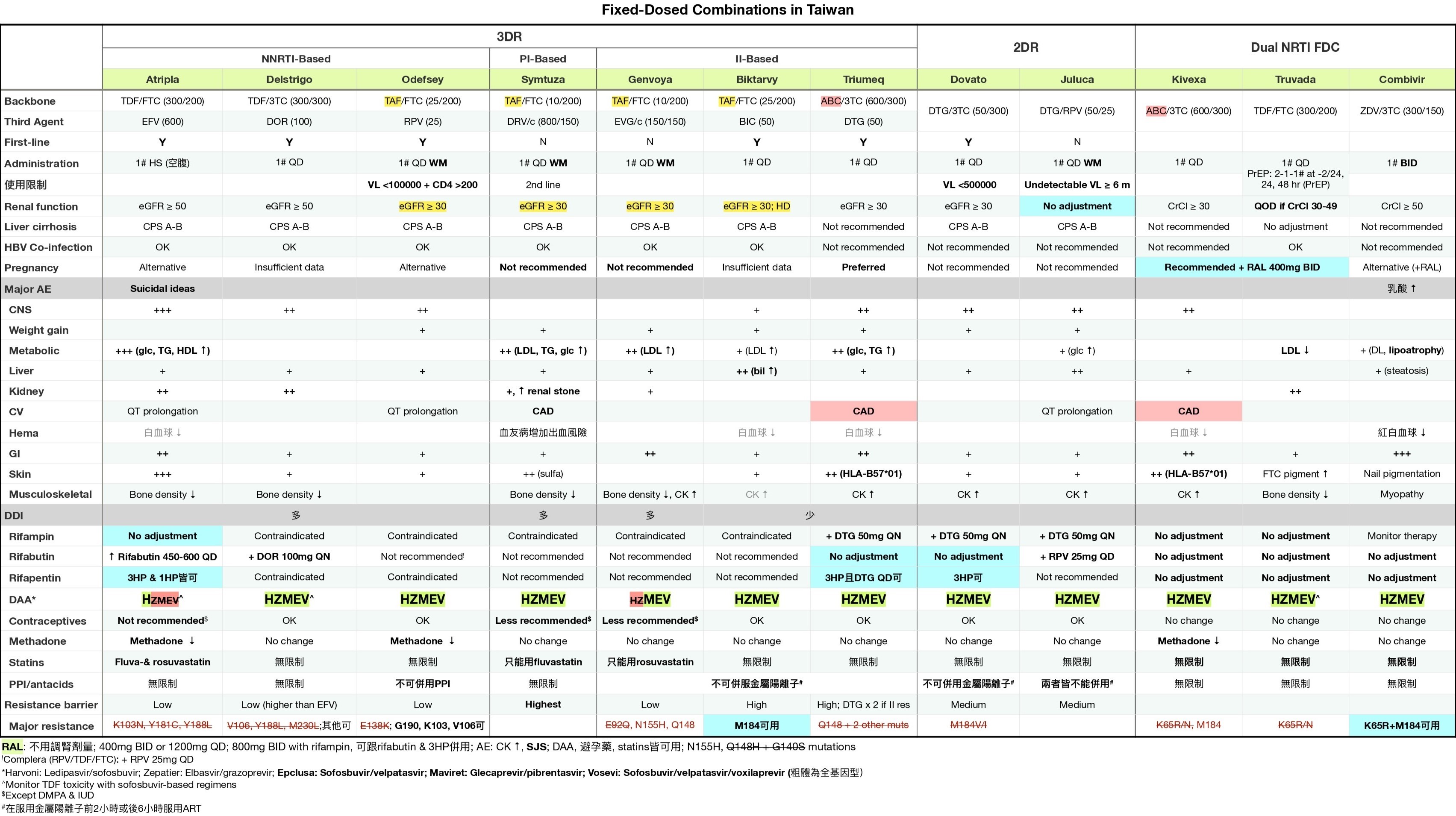

考古題

專師

// Specify the path to the original note
const originalNotePath = "Area/Medicine/專師考試解題.md";  // Replace with your actual file path

// Read the content of the original file asynchronously
const content = await app.vault.read(app.vault.getAbstractFileByPath(originalNotePath));

// Define the tag you're searching for
const tag = "#medicine/HIV";  // Replace with your actual tag

// Split the content into sections based on the start of a new question (line with a number and a period)
const sections = content.split(/(?=\d+\.\s)/);

// Initialize an array to store the results
const results = [];

// Process each section
sections.forEach(section => {
    if (section.includes(tag)) {
        // Add the section to results if it contains the tag
        results.push(section.trim());
    }
});

// Output the captured sections
results.forEach(result => dv.paragraph(result));
內科醫師

// Specify the path to the original note
const originalNotePath = "Area/Medicine/內專考古題.md";  // Replace with your actual file path

// Read the content of the original file asynchronously
const content = await app.vault.read(app.vault.getAbstractFileByPath(originalNotePath));

// Define the tag you're searching for
const tag = "#medicine/HIV";  // Replace with your actual tag

// Split the content into sections based on the start of a new question (line with a number and a period)
const sections = content.split(/(?=\d+\.\s)/);

// Initialize an array to store the results
const results = [];

// Process each section
sections.forEach(section => {
    if (section.includes(tag)) {
        // Add the section to results if it contains the tag
        results.push(section.trim());
    }
});

// Output the captured sections
results.forEach(result => dv.paragraph(result));
肺炎

up:: 專師考試解題

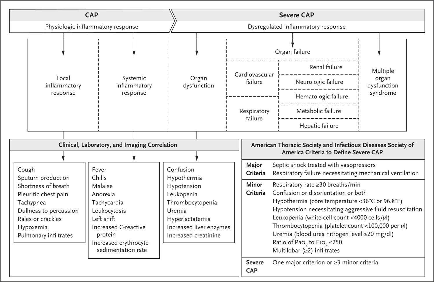
台灣肺炎診治指引

Definitions and clinical manifestations

  • Pneumonia: s/s (fever, cough, purulent sputum, dyspnea) + new infiltrate on chest imaging
  • Community-acquired pneumonia (CAP): pneumonia acquired outside of hospital setting
  • Hospital-acquired pneumonia (HAP): pneumonia acquired ≥48 hrs after hospitalization
  • Ventilator-associated pneumonia (VAP): pneumonia acquired ≥48 hrs after intubation
  • Lung empyema: accumulation of pus in pleural space
  • Lung abscess: parenchymal necrosis with confined cavitation
  • Aspiration pneumonitis: acute lung injury after inhalation of gastric contents without infection, though bacterial infection can occur within 24–72 hrs of injury

Microbiology of Pneumonia

Clinical Setting Etiologies
CAP (AJRCC 2019;200:7) No pathogen identified in 50–60%, virus alone in ~25%, bacteria alone in ~10%, virus-bacteria coinfection in <5%
Viruses: influenza, RSV, hMPV, parainfluenza, rhinovirus, coronavirus
S. pneumoniae (most common bacterial cause)
S. aureus (espec. post-influenza)
Mycoplasma, Chlamydia (espec. in young & healthy)
H. influenzae, M. catarrhalis (espec. in COPD)
Legionella (espec. in elderly, smokers, ↓ immunity, TNF inhibitors)
Klebsiella & other GNR (espec. in alcoholics & aspiration)
HAP/VAP S. aureus, Pseudomonas, Klebsiella, E. coli, Enterobacter, Acinetobacter, Steno. IV abx w/in 90 d risk factor for MDR. Viral ~20% cases.
Empyema S. pneumo, S. aureus, E. coli, Klebsiella, H. influenzae, anaerobes
Lung abscess Often polymicrobial, incl. oral flora. S. aureus, anaerobes, Strep (anginosus, GAS), GNR (Klebsiella, E. coli, Pseudomonas), Nocardia, Actinomyces, fungi, mycobacteria, Echinococcus
Immunosupp. Above + Pneumocystis, Cryptococcus, Nocardia, non-TB myco- bacteria (NTM), CMV, invasive molds
Pathogen Group Pathogen
Common or core
Gram-positive bacteria Streptococcus pneumoniae, methicillin-susceptible Staphylococcus aureus, Strep. pyogenes, other streptococci
Gram-negative bacteria Hemophilus influenzae, Moraxella catarrhalis, Enterobacteriaceae (e.g., Klebsiella pneumoniae)
Atypical bacteria Legionella pneumophila, Mycoplasma pneumoniae, Chlamydophila pneumoniae
Respiratory viruses Influenza virus, SARS-CoV-2, respiratory syncytial virus, parainfluenza virus, human metapneumovirus, rhinoviruses, common human coronaviruses
Uncommon or infrequent
Gram-positive bacteria Methicillin-resistant Staph. aureus, nocardia species, Rhodococcus equi
Gram-negative bacteria Enterobacteriaceae, including extended-spectrum beta-lactamases or carbapenem-resistant enterobacteriaceae; nonfermenting bacilli (e.g., pseudomonas or acinetobacter); Francisella tularensis
Atypical bacteria Chlamydia psittaci, Coxiella burnetii
Mycobacteria Mycobacterium tuberculosis, nontuberculous mycobacteria
Viruses Cytomegalovirus, herpes simplex, varicella zoster, MERS-CoV
Fungi Pneumocystis jirovecii, aspergillus species, mucorales species, histoplasma species, cryptococcus species, blastomyces species, coccidioides species
Parasites Strongyloides stercoralis, Toxoplasma gondii
CAP Pathogen Specific Risk factors
Influenza Influenza activity in the community. Close contact with an infected person.
SARS-CoV-2 SARS-CoV-2 activity in the community. Close contact with an infected person.
Legionella species Recent cruise ship. Exposure to contaminated water sources (e.g., hot tubs, cooling towers; recent plumbing)
MRSA Prior infection or colonization with MRSA
Pseudomonas aeruginosa Prior infection or colonization withPseudomonas aeruginosa
MDR Gram-negative rods Prior infection or colonization with MDR Gram-negative rods
Oral anaerobic bacteria Poor dental hygiene
Chlamydophila psittaci Exposure to birds
Coxiella burnetti Exposure to farm animals or parturient cats
Francisella tularensis Exposure to rabbits
MERS-CoV Exposure to infected camels; travel to the Arabian Peninsula
Coccidioides species Travel to southwestern United States
Histoplasma capsulatum Exposure to bat or bird droppings

Diagnostic studies

(AJRCC 2019;200:e45)

  • Sputum Gram stain/Cx:
    • Reliable if high quality (ie, sputum not saliva; <10 squam cells/lpf).
    • If bacterial PNA should be purulent (>25 PMN/lpf).
    • Yield ↓ >10 h after abx (CID 2014;58:1782).
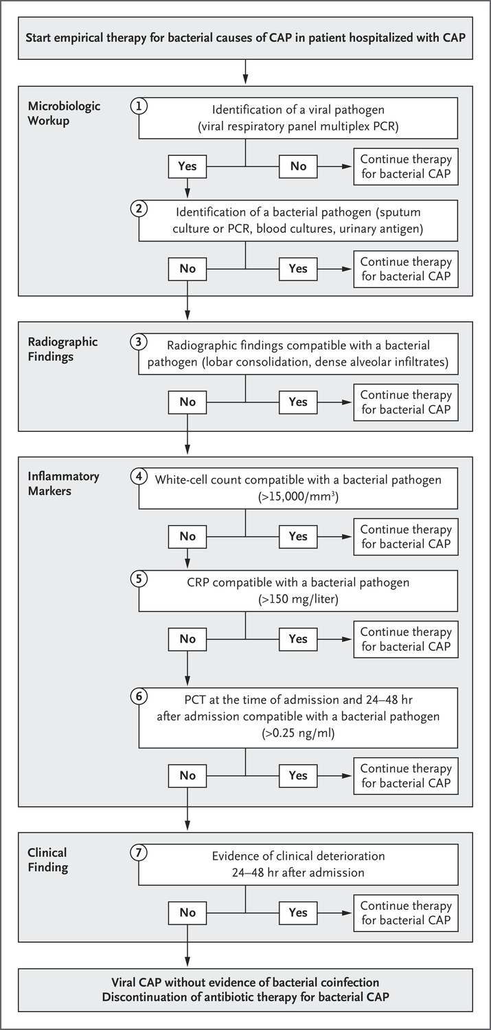
  • Procalcitonin: ↑ in acute bacterial (not viral) PNA.
    • Consider stopping abx if levels <0.25 ng/ml (<0.5 ng/mL in ICU Pts) or ↓ ≥80% from peak. ↓ abx exposure by 2–3 d (Lancet ID 2016;16:819 & 2018;18:95).
    • Not validated in immunosuppressed hosts.
    • Levels harder to interpret in CKD.
    • False ⊕ in cardiac arrest, shock, burns, surgery.
  • CXR (PA & lateral)
  • HIV test (if unknown)
  • MRSA nares swab in HAP/VAP (if ⊖ 96% NPV for MRSA PNA)
  • Consider in severe disease (otherwise not recommended):
    • Legionella urinary Ag (detects L. pneumophila L1 serotype, 60–70% of clinical disease)
    • S. pneumoniae urinary Ag (Se 70%, Sp >90%)
    • Blood cultures (before antibiotics!): ⊕ in ~10% of inPts, depending on pathogen
  • If clinical suspicion for mTB:
    • (induced) sputum AFB stain ×3 q ≥8h (w/ ≥1 early morning).
    • Mycobact. cx (empiric respiratory isolation while pending)
    • MTb DNA PCR if smear ⊕
  • Viral testing (DFA or PCR) on nasopharyngeal swab or sputum
  • Bronchoscopy: immunosupp., critically ill, failure to respond, suspected PCP, inadequate/ ⊖ sputum cx
    • Send Gram stain/cx, Legionella cx, fungal cx/wet prep, mycobacterial cx/smear, modified AFB stain, galactomannan
  • Reasons for failure to improve on initial treatment:
    • Insufficient time: may take ≥72 h to see improvement (fever persists >4 d in ~20%)
    • Insufficient drug levels for lung penetration (eg, vanco trough <15–20 μg/mL)
    • Resistant organisms or superinfxn: eg, MRSA, Pseudo.; consider bronchoscopy
    • Wrong dx: fungal/viral, chemical pneumonitis, PE, CHF, ARDS, DAH, ILD; consider CT
    • Parapneumonic effusion/empyema/abscess: if CXR ⊖, consider bedside US or CT. If effusion >1 cm, drain & send fluid pH, gluc, Gram stain & Cx.
    • Metastatic infection (eg, endocarditis, septic arthritis)

Triage

  • qSOFA predicts poor outcomes, prolonged ICU stay, and in-hospital mortality if >2 of 3: RR>22, AMS, SBP<100 (JAMA 2016; 315:801)

Treatment

(NEJM 2019;380:651; AJRCC 2019;200:e45)

  • Avoid quinolones if suspect TB. When possible, de-escalate abx based on sensitivities.
  • Steroids: not unless indicated for shock or COPD exacerbation; may ↓ mortality, mech vent, & ARDS in severe CAP (Cochrane 2017;12:CD007720). Avoid in influenza.
  • Duration:
    • CAP: 5–7 days, can de-escalate IV abx to PO after clinical improvement.
    • HAP/VAP: 7 days.
    • Empyema/abscess: 2–6 wks based on complexity, drainage.
Scenario Regimen
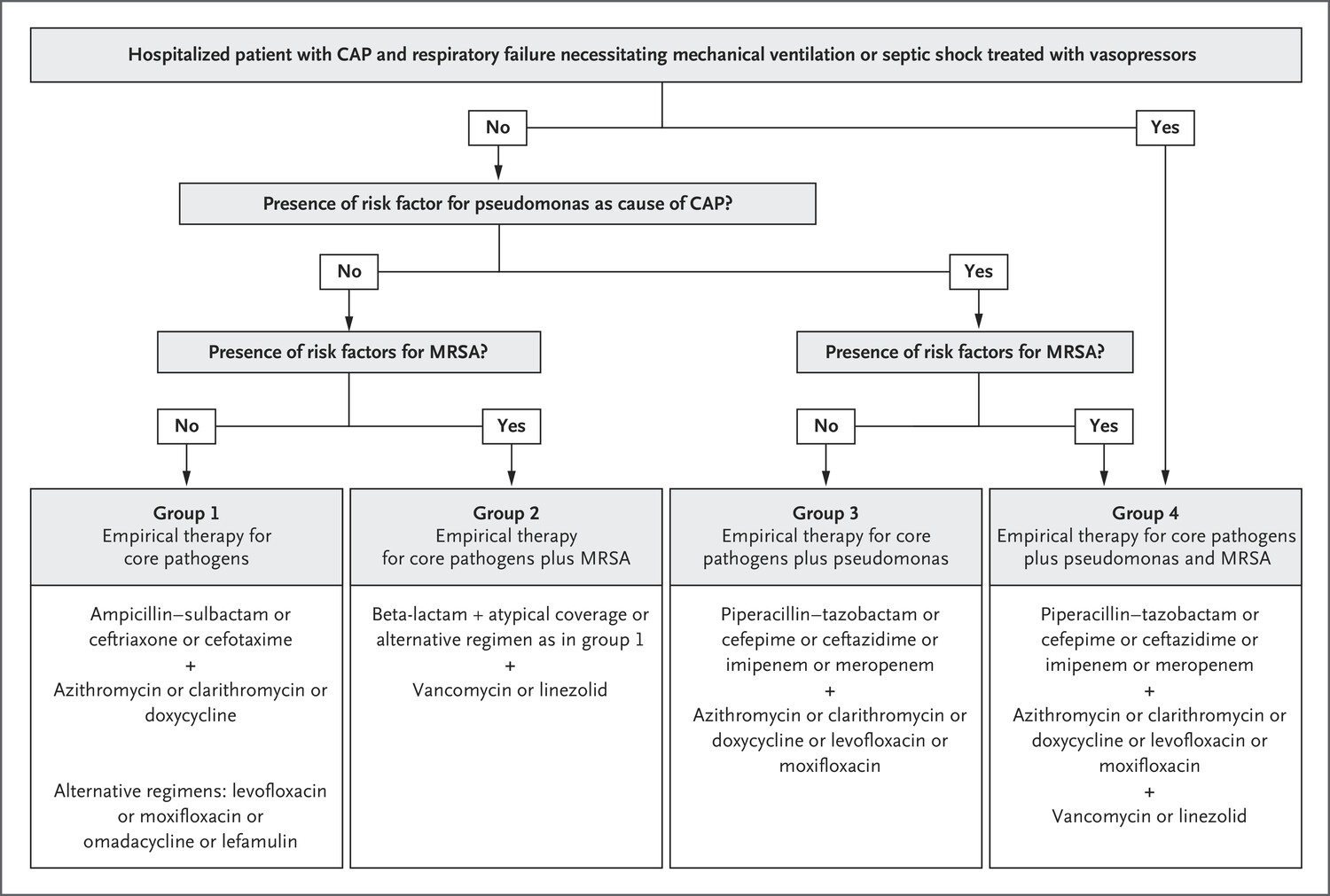
CAP (outPt) Amoxicillin, azithro, or doxy (avoid latter two if >25% resistance locally)
CAP (ward) [3rd-gen ceph + azithro] or levoflox; omadacycline ≍ FQ (NEJM 2019;380:517)
CAP (ICU) 3rd-gen ceph + azithro. Only cover MRSA or Pseudomonas if risk factors (prior PsA PNA, MRSA infection, recent hospitalization, IV abx)
HAP/VAP [Pip-tazo or cefepime or carbapen.] + [vanc or linezolid]. May add resp FQ or azithro if concerned for atypicals. Daptomycin not active in lungs.
Empyema/ abscess [3rd-gen ceph + MNZ] or amp-sulbactam. Only cover Pseudomonas or MRSA if risk factors. Empyema: drain if >1 cm ± chest tube. Abscess: drainage not required. De-escalate to PO abx based on clinical improvement & micro.


Prevention

  • All persons >65 or age 19–64 w/ CHF, lung disease, cirrhosis, DM, EtOH, smoker, immunosupp. (eg, ESRD, organ transplant, HIV, leukemia, lymphoma, asplenia)
  • PCV20 vaccine or PCV15 + PPSV23 1 yr later

Viral Respiratory Infections

Presentation: URI, bronchitis, bronchiolitis, pneumonia (Lancet 2011;377:1264)

Microbiology & epidemiology

(http://www.cdc.gov/flu/weekly

  • Typical pathogens: Short, mild = rhinovirus, other non-SARS-CoV-2 coronavirus.
  • Longer, more severe or complicated = influenza, parainfluenza, RSV, adenovirus, metapneumovirus, COVID-19 (vide infra). Can be esp. severe in immunosupp.

Diagnosis

  • Sx: fever, cough, myalgias, SOB, wheezing, sore throat, rhinorrhea, malaise, confusion
  • Respiratory viral panel on nasal swab or sputum/BAL; rapid flu nasopharyngeal swab preferred to nasal swab (Se 50–70%, Sp >90%); RT-PCR for flu A/B (>95% Se & Sp)

Treatment

(NEJM 2017;390:697) 

  • Influenza (A & B):
    • Neuraminidase inhib. (eg, oseltamivir); must start w/in 48 h of sx for low-risk; for critically ill or immunosupp., start ASAP even if >48 h. Peramivir IV if unable to tolerate PO.
    • Endonuclease inhib. (baloxavir): superior to oseltamivir in ↓ sx & viral load on 1st day of Rx, but resistance emerging; no data in severe influenza (NEJM 2018; 379:913)
  • RSV: can consider inhaled ribavirin in immunosupp, but very expensive & rarely used

Prevention

  • Inactivated influenza vaccine: rec for all >6 mo of age.
  • Isolation, droplet precautions for inpatients strongly recommended
  • Ppx for high-risk contacts of confirmed influenza: oseltamivir × 7 d or baloxavir single dose

考古題

專師

// Specify the path to the original note
const originalNotePath = "Area/Medicine/專師考試解題.md";  // Replace with your actual file path

// Read the content of the original file asynchronously
const content = await app.vault.read(app.vault.getAbstractFileByPath(originalNotePath));

// Define the tag you're searching for
const tag = "#medicine/pneumonia";  // Replace with your actual tag

// Split the content into sections based on the start of a new question (line with a number and a period)
const sections = content.split(/(?=\d+\.\s)/);

// Initialize an array to store the results
const results = [];

// Process each section
sections.forEach(section => {
    if (section.includes(tag)) {
        // Add the section to results if it contains the tag
        results.push(section.trim());
    }
});

// Output the captured sections
results.forEach(result => dv.paragraph(result));
內科醫師

// Specify the path to the original note
const originalNotePath = "Area/Medicine/內專考古題.md";  // Replace with your actual file path

// Read the content of the original file asynchronously
const content = await app.vault.read(app.vault.getAbstractFileByPath(originalNotePath));

// Define the tag you're searching for
const tag = "#medicine/pneumonia";  // Replace with your actual tag

// Split the content into sections based on the start of a new question (line with a number and a period)
const sections = content.split(/(?=\d+\.\s)/);

// Initialize an array to store the results
const results = [];

// Process each section
sections.forEach(section => {
    if (section.includes(tag)) {
        // Add the section to results if it contains the tag
        results.push(section.trim());
    }
});

// Output the captured sections
results.forEach(result => dv.paragraph(result));
皮膚與軟組織感染

Source: Pocket Medicine, 2022

up:: 專師考試解題

定義

  • 蜂窩性組織炎:是真皮和皮下組織的感染,特徵為紅斑、發熱、壓痛和腫脹;通常因皮膚破損引起(JAMA 2016;315:3)。
  • 皮膚膿瘍:皮下膿液的聚集。
  • 葡萄球菌中毒性休克症候群(Staphylococcal toxic shock syndrome):快速發作的發燒、皮疹、低血壓和多重器官損傷。診斷不需要葡萄球菌的培養。通常與填充物(如衛生棉條、鼻腔填充物)有關。處理可能需要手術清創加上抗生素治療。

風險因素

  • 創傷、水腫、先前的皮膚炎症或感染、肥胖、糖尿病、其他免疫抑制情況(CID 2014;59:e10)。

微生物學

  • 化膿性:MRSA(NEJM 2006;355:666)引起多達75%的化膿性皮膚/軟組織感染,其次是MSSA和鏈球菌。
  • 非化膿性:鏈球菌、MSSA、需氧革蘭氏陰性桿菌。MRSA較不常見,除非有顯著風險因素(如先前MRSA感染、靜脈注射藥物使用者、血液透析、近期使用抗生素或住院)。
  • 咬傷:皮膚(鏈球菌、葡萄球菌 [僅在有風險因素時考慮MRSA])和口腔菌群(包括厭氧菌)加上特殊暴露:
特徵 微生物學 臨床表現 治療
貓咬傷^ Pasturella spp 快速出現紅斑、腫脹、淋巴管炎、發燒 Amox/clav
狗咬傷 Pasturella & Capnocytophaga spp 在無脾患者/肝硬化患者和其他免疫抑制患者中可引起嚴重敗血症,伴隨DIC和壞疽 Amox/clav

如懷疑感染Capno.:使用pip/tazo或carbapenem
穿透性損傷 Pseudomonas 可能與深部組織膿瘍有關 根據敏感性選擇治療方案
園藝活動 Sporothrix 潰瘍性結節,延淋巴擴散 Itraconazole
鹽水或生蠔/魚暴露 V. vulnificus 出血性水疱及敗血症(尤其在肝硬化患者中) Doxy + Ceftazidime/ceftriaxone
Mycobacterium marinum 緩慢進展,四肢結節/淺表淋巴結炎 Macrolide+rifampin/ethambutol
淡水暴露 Aeromonas 可導致肌壞死/橫紋肌溶解 FQ, TMP-SMX, 或 ceftriaxone
^貓抓病由Bartonella通過貓抓或咬傷引起。導致淋巴結炎。

診斷

  • 根據理學檢查進行臨床診斷
  • 不應從完整皮膚上進行培養,因為沒有幫助(CID 2014;59:e10)
  • 血液培養通常陽性率很低(約5-10%)
  • 超音波檢查可用於識別深部膿瘍並幫助引流。如果發現膿瘍,切開引流是治療的關鍵。
  • 膿瘍抽取物可能提供微生物學診斷

治療(NEJM 2014;370:2238; CID 2014;59:e10; JAMA 2016;316:325 & 2017;317:2088)

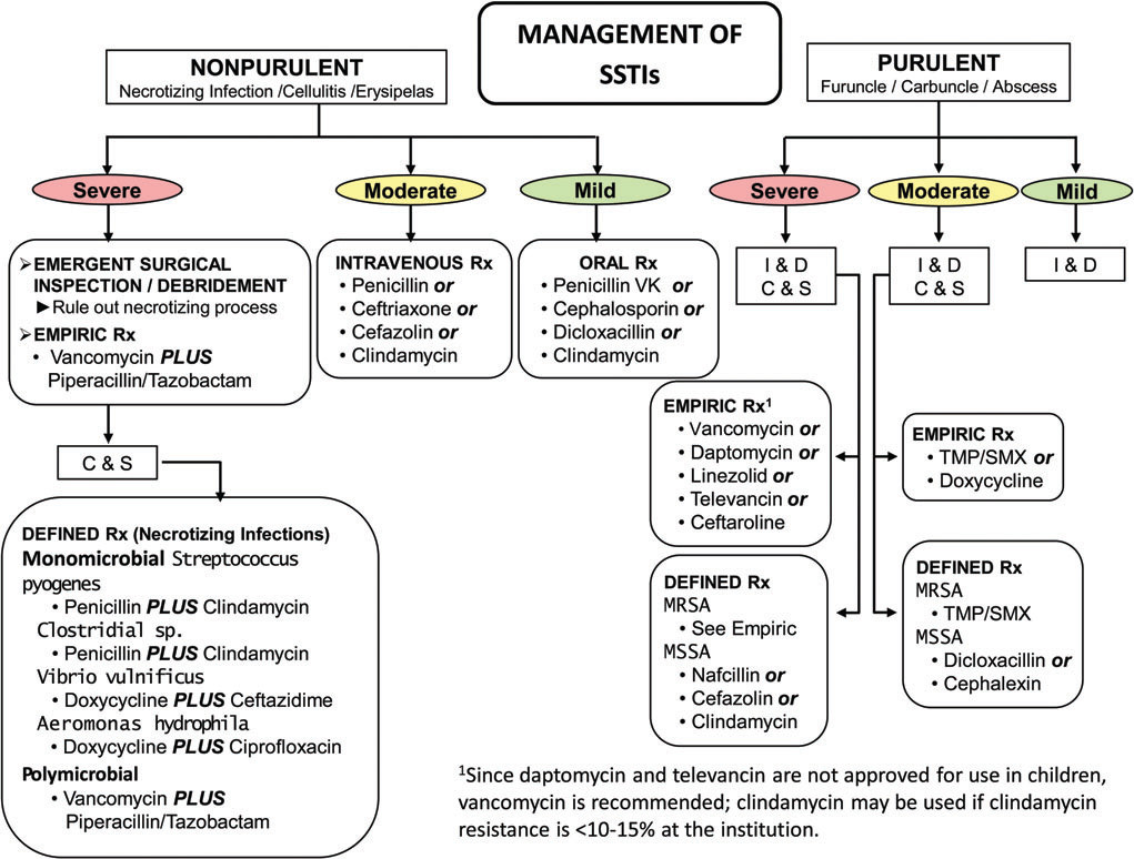
是否化膿性 常見的微生物 嚴重程度 治療
β-溶血性鏈球菌 > S. aureus  輕度 口服:penicillin VK、cephalosporin
中度 靜脈注射:penicillin、ceftriaxone、cefazolin
重度 靜脈注射:vanc + pip/tazo(± clinda if TSS)
S. aureus(包括MRSA)>> β-溶血性鏈球菌 輕度 I&D only 或 I&D + clinda or TMP-SMX(NEJM 2017;376:2545)
中度 I&D + TMP-SMX or doxycycline
重度 I&D + 靜脈注射vancomycin、daptomycin或linezolid(± clinda if TSS)

輕度:膿瘍小於2公分,無全身性感染症狀,免疫功能正常,無植入物;中度:全身性症狀;重度:SIRS或免疫抑制

  • 抬高患肢;開始使用抗生素後紅斑可能加重,因為細菌殺死後會導致發炎
  • 在肥胖患者中,為避免治療失敗,應確保足夠的藥物劑量(J Infect 2012;2:128)
  • 治療持續時間:根據嚴重程度和治療反應,從5天到最多14天。拍照並畫出邊界以追蹤進展

壞死性軟組織感染(NEJM 2017;377:2253)

定義

  • 猛爆性組織破壞、全身性毒性和高死亡率。屬於外科急症
  • 可能包括蜂窩性組織炎、筋膜炎、肌炎、肌壞死(產氣性壞疽)

風險因素

  • 通常通過皮膚/黏膜損傷或創傷性傷口影響健康人,但糖尿病、周邊血管疾病、酗酒、靜脈注射藥物使用者、肝硬化或其他免疫抑制者風險增加

微生物學

壞死性筋膜炎

  • 第一型:多重病原菌(混合嗜氧菌和厭氧菌),通常發生於具有上述風險因素的老年患者
    • Fournier氏壞疽影響生殖器和/或會陰部
    • 頭頸部壞死性軟組織感染由口腔菌群(包括厭氧菌)引起
  • 第二型:單一病原菌,通常為A群鏈球菌,較少見的是S. aureusVibrioAeromonas;與TSS相關
  • Clostridial myonecrosis(氣性壞疽):由C. perfringensC. septicum引起,革蘭氏染色顯示大革蘭氏陽性桿菌,端部鈍圓。與創傷性傷口相關,這些創傷性傷口創造了適合梭狀芽孢桿菌生長的厭氧環境

臨床表現

  • 紅斑、水腫、發熱加上全身性症狀,可能伴隨有捻髮感、水疱、壞死。
  • 臨床症狀迅速惡化
  • 疼痛程度超過明顯的蜂窩性組織炎;皮膚先出現感覺過敏,後來則感覺喪失

診斷

  • 臨床診斷足以開始緊急手術探查
  • 壞死中心的抽吸物;血液培養;革蘭氏染色;乳酸、AST和CK以檢查深部組織壞死
  • 影像學檢查:無顯影劑的CT檢查,但不應延遲治療/手術(Arch Surg 2010;145:452)
  • 手術檢體的革蘭氏染色和培養可進行微生物學診斷

治療(CID 2014;60:169)

  • 緊急手術探查和壞死組織的清創,並諮詢感染科
  • 經驗性抗生素: [pip/tazo or (ceftriaxone + metronidazole) or carbapenem] + [vancomycin或linezolid]。
  • A群鏈球菌:penicillin + clindamycin,並考慮使用IVIG治療中毒性休克

糖尿病足部感染

微生物學與嚴重程度 (CID 2004;39:885)

  • 輕度(表淺性潰瘍,無深層結構受累,周圍紅斑 <2公分,且無全身性症狀):通常為S. aureus或嗜氧性鏈球菌
  • 中度(潰瘍伴有深層結構受累,周圍紅斑 >2公分,或淋巴管發炎性條紋且無全身性症狀):更可能為慢性及多重微生物感染(Pseudomonas aeruginosa、腸球菌、腸道革蘭氏陰性桿菌、厭氧菌)
  • 嚴重(中度 + 全身性症狀或代謝不穩定):厭氧性鏈球菌、腸道革蘭氏陰性桿菌、Pseudomonas aeruginosa、Bacteroides、Clostridium

初步評估

  • 清創、去除壞死組織、探查並取得深部厭氧菌及嗜氧菌培養
  • 評估周邊血管疾病:感覺、脈搏、踝臂指數(ABI)

診斷

  • 清創時取深層組織培養(理想情況下在使用抗生素前)。表面拭子通常用途有限,多為移生菌。
  • 中度/嚴重感染:取得血液培養、紅血球沉降率(ESR)、C反應蛋白(CRP)
  • 應排除骨髓炎。如果有以下情況風險較高:骨骼明顯可見或可探查到骨骼、潰瘍 >2 公分、潰瘍持續時間 >1-2 週、ESR >70。如果懷疑骨髓炎,應取得X光或核磁共振影像(見下方骨髓炎部分)。

治療 (CID 2012;54:e132)

  • 輕度感染:口服抗生素。針對革蘭氏陽性菌(dicloxacillin、cephalexin 或 amox/clav);使用 TMP-SMX 或 doxy 對付 MRSA。
  • 中度/嚴重感染:靜脈注射抗生素。針對革蘭氏陽性菌(vancomycin、linezolid、daptomycin) + 革蘭氏陰性菌(ceftriaxone、levofloxacin 或 amp/sulb)± 厭氧菌(metronidazole 或 clindamycin)。如果有以下情況,增加抗Pseudomonas aeruginosa抗生素(cefepime 或 pip/tazo):浸軟傷口、顯著水域暴露、溫暖氣候
  • 抬高患肢,避免負重,傷口護理,控制血糖,治療靜脈功能不全和動脈缺血
  • 許多需要手術:早期、積極且重複的清創;可能需要血管重建或截肢

骨髓炎

  • 骨骼感染,可能是由血源性感染或直接從鄰近部位擴散

病因 (Lancet 2004;364:369)

  • 血源性感染:S. aureus;脊椎體結核感染 = Pott 病
  • 鄰近部位感染(可能是急性或慢性)
  • 開放性骨折、骨科手術等:S. aureusS. epidermidis (coagulase-negative Staphylococcus)
  • 皮膚破損 + 血管功能不全(例如糖尿病足、壓瘡):多重微生物感染
  • 泌尿系統源(革蘭氏陰性桿菌、腸球菌)

臨床表現

  • 周圍軟組織受損 ± 瘻管至表面皮膚
  • ± 發燒、倦怠和夜間盜汗(血源性感染較常見)
  • 脊椎骨髓炎(尤其是靜脈注射毒品者):持續性局部背痛,± 發燒(NEJM 2010;362:1022)

診斷 (JAMA 2008;299:806)

  • 確保取得病原體培養數據,以避免長期使用經驗性抗生素
  • 骨組織切片或組織培養,透過手術或經皮切片獲得(送嗜氧菌、厭氧菌、結核菌和真菌培養 + 病理檢查),除非血液培養結果為陽性。不要依賴潰瘍或瘻管引流的拭子。
  • 理學檢查:如果在糖尿病足(見上方)中能探查潰瘍至骨骼潰瘍 >2 cm²,則高度懷疑(靈敏度 83%,特異度 90%)
  • 使用抗生素前進行血液培養(急性血源性骨髓炎更常陽性)
  • CBC、CRP、ESR(>70 大幅增加骨髓炎的可能性)
  • 影像學檢查
  • 普通 X 光片:早期疾病正常;2-6 週後可見溶骨性病變
  • 磁共振影像(MRI):首選影像學檢查(整體靈敏度 90%,特異度 82%;Archives 2007;167:125)
  • CT:可以顯示骨膜反應及皮質和骨髓破壞
  • CT 和 MRI 雖靈敏度高但特異度低;如果是鄰近焦點伴骨膜反應或 Charcot 改變,可能會偽陽性
  • 核醫影像:靈敏度高但特異度低(如果有軟組織發炎可能偽陽性)

治療

  • 抗生素:根據培養數據。如果臨床穩定,考慮在取得骨切片結果前暫時停止使用抗生素。治療期間取決於治療策略/治療目標(例如,脊椎骨髓炎 6 週;Lancet 2015;385:875)。在靜脈抗生素或手術後 ≥7 天,如果情況良好,考慮(在與感染科醫師商討後)將靜脈改為口服(如果口服抗生素有良好的生物利用度和骨髓穿透能力)(NEJM 2019;380:425)。
  • 對於以下任何情況,應考慮手術:急性骨髓炎對醫療治療無效、慢性骨髓炎、化膿性脊椎骨髓炎的併發症(例如神經功能損害、脊椎不穩定、硬膜外膿腫)或感染的人工植入物

%%Original: SSTI%%

考古題

專師

// Specify the path to the original note
const originalNotePath = "Area/Medicine/專師考試解題.md";  // Replace with your actual file path

// Read the content of the original file asynchronously
const content = await app.vault.read(app.vault.getAbstractFileByPath(originalNotePath));

// Define the tag you're searching for
const tag = "#medicine/SSTI";  // Replace with your actual tag

// Split the content into sections based on the start of a new question (line with a number and a period)
const sections = content.split(/(?=\d+\.\s)/);

// Initialize an array to store the results
const results = [];

// Process each section
sections.forEach(section => {
    if (section.includes(tag)) {
        // Add the section to results if it contains the tag
        results.push(section.trim());
    }
});

// Output the captured sections
results.forEach(result => dv.paragraph(result));

內科

// Specify the path to the original note
const originalNotePath = "Area/Medicine/內專考古題.md";  // Replace with your actual file path

// Read the content of the original file asynchronously
const content = await app.vault.read(app.vault.getAbstractFileByPath(originalNotePath));

// Define the tag you're searching for
const tag = "#medicine/SSTI";  // Replace with your actual tag

// Split the content into sections based on the start of a new question (line with a number and a period)
const sections = content.split(/(?=\d+\.\s)/);

// Initialize an array to store the results
const results = [];

// Process each section
sections.forEach(section => {
    if (section.includes(tag)) {
        // Add the section to results if it contains the tag
        results.push(section.trim());
    }
});

// Output the captured sections
results.forEach(result => dv.paragraph(result));

敗血症與休克

Source: Pocket Medicine, 2022

up:: 專師考試解題

休克

  • 組織缺氧,因為組織灌流降低,進而降低組織氧氣供應 和/或 增加氧氣消耗 或 氧氣利用不當
  • 典型的體徵包括低血壓(收縮壓 <90 mmHg 或收縮壓下降 >40 mmHg)、心搏過速、少尿(尿量 <0.5 cc/kg/h)、意識變化、代謝性酸中毒 ± 乳酸增加
  • 診斷困難,因為全身性血管阻力(SVR)增加可能維持收縮壓,但組織灌流差;休克指數(心率/收縮壓)>0.9脈壓[(收縮壓 - 舒張壓)/收縮壓] <25% 是顯著休克的線索

各種休克形式的血流動力學特徵 (NEJM 2013;369:1726)

休克類型 右心房 肺微血管楔壓 心輸出量 全身血管阻力
低血容量性休克
心因性休克 正常或 ↑
右心室梗塞/大範圍肺栓塞 正常或 ↓
心包填塞
分布性休克 變化不定 變化不定 通常 ↑(在敗血症中可能 ↓)

替代指標:右心房壓 ≈ 頸靜脈壓(1 mmHg = 1.36 cm H2O);胸部X光顯示肺水腫暗示肺微血管楔壓 (PCWP) ↑;尿量 ∝ 心排出量(排除急性腎損傷);延遲毛細血管充盈時間(即 >2-3秒)暗示系統性血管阻力 ↑

定義

系統性發炎反應症候群 (SIRS)

定義為符合以下任==兩項==標準:
- 體溫高於 38°C 或低於 36°C
- 心跳超過 90 次/分鐘
- 呼吸速率超過 20 次/分鐘 或 二氧化碳分壓低於 32 mmHg
- 白血球數量超過 12000 或低於 4000 /微升,或未成熟細胞或帶狀核白血球超過 10%

敗血症

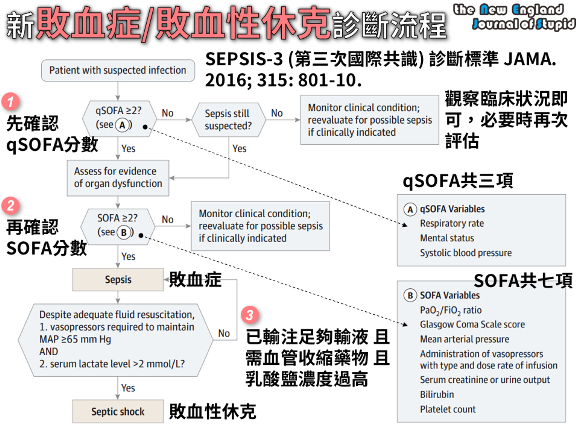
  • 由感染引起的具生命威脅器官功能障礙(SOFA ≥2)
  • ==qSOFA:符合呼吸頻率 ≥22,意識變化,收縮壓 ≤100 mmHg==任兩項或以上

敗血性休克

  • 敗血症引起的循環和細胞/代謝異常,足以增加死亡率;即使在適當的液體復甦後,仍==需使用升壓劑以使平均動脈壓(MAP) ≥65且乳酸 >2==
  • Sequential Organ Failure Assessment (SOFA):呼吸(P/F ratio降低);凝血(==血小板==降低);肝臟(膽紅素升高);心血管(MAP下降或升壓劑增加);中樞神經系統(GCS 降低);腎臟(肌酐升高或尿量下降)


Source: NEJS

處置 (Crit Care Med 2021;49:e1063)

液體

  • 在發病後3小時內進行積極的靜脈液體復甦(30 mL/kg),以bolus劑量給藥
  • 晶體溶液在復甦效果上與膠體溶液相當(JAMA 2013;310:1809; NEJM 2014;370:1412);不考慮使用明膠(gelatin)復甦 (CritCareMed 2021;49:e1063)
  • 平衡晶體液(乳酸林格液、Plasma-Lyte)與生理鹽水在死亡率、器官衰竭或需要腎替代療法方面未顯示一致的好處(NEJM 2018;378:829 & 2022:386:815)
  • NaHCO3可能在 急性腎損傷及pH <7.2 的情況下降低死亡率和對腎替代療法的需求(Lancet 2018;392:31)
  • 液體反應性預測因子:脈壓變化 >13% 與呼吸(Chest 2008;133:252);靜脈腔(IVC)直徑的呼吸變化,或在被動抬腿時脈壓上升 >10%。靜態中心靜脈壓(CVP)是較差的替代指標。
  • 在早期復甦後,如果出現急性肺損傷/急性呼吸窘迫綜合症(ALI/ARDS),目標中心靜脈壓(CVP)為4-6 mmHg,因為額外的液體可能有害 → 增加呼吸器/ICU天數(NEJM 2006;354:2564; Chest 2008;133:252)

升壓劑與強心劑

  • 目標MAP 65 mmHg 與 80-85 mmHg 一樣好,且可減少心房顫動(NEJM 2014;370:1583; JAMA 2020;323:938)
  • Norepinephrine:相比於dopamine 減少心律失常與死亡率(NEJM 2010;362:779; Crit Care Med 2012;40:725),並且是敗血性休克的首選升壓劑
  • Vasopressin:添加至 norepinephrine(而不是使用高劑量 norepinephrine)可減少心房顫動與腎替代療法的風險約¼(JAMA 2018;319:1889)
  • 若為難治性血管舒張性休克(refractory vasoplegia):可使用血管張力素 II(Giaprezza)、亞甲藍(methylene blue)、類固醇(見下文)
  • 若在適當的液體與升壓劑治療後未達目標(見下文),考慮使用強心劑

目標

  • 乳酸清除率(≥20%/2 小時)與ScvO2 指導復甦效果一樣好(JAMA 2010;303:739)
  • 目標==毛細血管充盈時間 ≤3秒==(每30分鐘檢查)與乳酸清除率效果相當或更好(JAMA 2019;321:654)

抗生素

  • 在確認重症敗血症或敗血性休克後,儘快開始經靜脈抗生素治療;每小時延遲抗生素給藥與死亡率增加7.6% 相關(Crit Care Med 2006;34:1589),在急診部門發病後3小時內給予抗生素與降低住院死亡率相關(NEJM 2017;376:2235)
  • 很可能敗血症或敗血性休克成人,建議==1小時內==投與抗生素;可能敗血症(但非休克)成人,考慮==3小時內==投與抗生素;感染低風險且沒有休克成人,考慮延後使用抗生素、緊密觀察 (CritCareMed 2021;49:e1063)
  • 如有可能,在緊急開始抗生素前獲取2組血液培養(但不要延遲抗生素給藥)
  • 廣效抗革蘭氏陽性菌(包括MRSA)與革蘭氏陰性菌(包括高度耐藥菌)覆蓋,±厭氧菌
  • 以降鈣素原為指導的抗生素==停用==(而非啟動)可降低死亡率(Crit Care Med 2018;46:684)

類固醇

  • Hydrocortisone 50 mg 靜脈注射每6小時一次 + fludrocortisone 50 μg 透過鼻胃管每日一次,可縮短休克持續時間並可能降低死亡率(NEJM 2018; 378:797 & 809)
  • 敗血性休克成人且有持續使用血管收縮藥物需求,考慮使用IV類固醇 (CritCareMed 2021;49:e1063)

早期目標導向治療 (EGDT)

  • 歷史上:在==6小時內使用液體與升壓劑達到MAP ≥65 mmHg,CVP 8-12 mmHg,尿量 ≥0.5 mL/kg/h;使用強心劑與紅血球濃縮液達到 ==ScvO2 ≥70% (NEJM 2001;345:1368)
  • 然而,現在在早期抗生素和充分液體復甦的時代,EGDT相對於目前的常規護理並未降低死亡率,且增加醫院成本(NEJM 2017;376:2223)

其他

  • 敗血症引起的低血氧呼吸衰竭,考量使用高流量鼻氧(HFNC)優於非侵入性通氣 (CritCareMed 2021;49:e1063)
  • 血糖控制目標為最高不要超過==180 mg/dl==,最低不要低於==110 mg/dl==
  • 輸血小板時機:
    • 血小板 < 10,000,不管有無出血
    • 血小板 < 20,000,有出血風險
    • 血小板 < 50,000,有出血或執行侵入性處置
  • 紅血球輸血只在血紅素低於 7.0 g/dL 時進行,目標是將血紅素提高到 7.0 ~ 9.0 g/dL 之間

Original: Sepsis and Shock

考古題

專師

// Specify the path to the original note
const originalNotePath = "Area/Medicine/專師考試解題.md";  // Replace with your actual file path

// Read the content of the original file asynchronously
const content = await app.vault.read(app.vault.getAbstractFileByPath(originalNotePath));

// Define the tag you're searching for
const tag = "#medicine/sepsis";  // Replace with your actual tag

// Split the content into sections based on the start of a new question (line with a number and a period)
const sections = content.split(/(?=\d+\.\s)/);

// Initialize an array to store the results
const results = [];

// Process each section
sections.forEach(section => {
    if (section.includes(tag)) {
        // Add the section to results if it contains the tag
        results.push(section.trim());
    }
});

// Output the captured sections
results.forEach(result => dv.paragraph(result));

Urinary tract infection

Source: Pocket Medicine, 2022

up:: 專師考試解題

定義

  • 無症狀菌尿症:尿液中存在細菌但無感染的徵兆或症狀
  • 簡單性:侷限於膀胱,無上尿路或全身感染的跡象
  • 複雜性
    • 超越膀胱(如腎盂腎炎、腎/腎周膿腫、前列腺炎),伴隨發燒、寒戰、倦怠、側腹痛、肋脊角壓痛或骨盆/會陰痛等症狀
    • 更可能發展為菌血症或敗血症
    • 男性、患有腎結石、狹窄、支架、尿路改道、免疫抑制、糖尿病者已不自動歸類為複雜性
    • 孕婦腎移植患者視為複雜性

微生物學

  • 簡單性
    • Escherichia coli大腸桿菌(80%)
    • Proteus mirabilis變形桿菌:與結石相關
    • Klebsiella克雷伯氏菌
    • S. saprophyticus腐生葡萄球菌:與女性使用衛生棉條有關(CID 2004;39:75)
    • 在健康的非孕婦中,lactobacilli乳酸桿菌、enterococci腸球菌、group B Streptococcus和coagulase-negative Staphylococcus(除腐生葡萄球菌外)可能是污染物(Annals 2012;156:ITC3)
  • 複雜性:如上 + Pseudomonas aeruginosa綠膿桿菌、enterococci腸球菌、staphylococci葡萄球菌(若無導管或近期手術器械使用史則不常見;?可能有菌血症並經血行擴散)。多重抗藥性增加。
  • 導管相關:最常見的是Escherichia coli大腸桿菌、Candida念珠菌、enterococci腸球菌、Pseudomonas aeruginosa綠膿桿菌及其他革蘭氏陰性桿菌。
  • 尿道炎Chlamydia trachomatis沙眼披衣菌, Neisseria gonorrhoeae淋病雙球菌, Ureaplasma urealyticum, Trichomonas vaginalis陰道滴蟲, Mycoplasma genitalium, HSV單純皰疹病毒。

臨床表現

  • 膀胱炎:排尿困難、尿急、尿頻、血尿、恥骨上疼痛;無發燒。若有膀胱炎和尿道炎症狀,需排除陰道炎。神經性膀胱患者可能有非典型症狀(如痙攣增加、自主神經反射異常、不適)。
  • 尿道炎:排尿困難、尿道分泌物
  • 前列腺炎
    • 慢性:類似膀胱炎 + 梗阻症狀(如排尿困難、尿流弱)
    • 急性:會陰疼痛、發燒、前列腺檢查時疼痛
  • 腎盂腎炎:發燒、寒戰、側腹或背部疼痛、噁心、嘔吐、腹瀉
  • 腎膿腫:腎盂腎炎症狀 + 在使用適當抗生素後仍持續發燒

診斷(NEJM 2016;374:562)

  • 尿液分析:膿尿 + 菌尿 ± 血尿 ± 硝酸鹽
  • 尿培養(清潔中段尿或直接導尿):
    • 只有在有症狀時才進行培養(但在病情嚴重的患者中,可包括意識變化、自主神經不穩定)
    • 如果 ≥105 CFU/mL 為陽性,然而 <105 但 ≥102/mL 在某些情況下仍可能表明尿路感染
    • 無菌性膿尿:尿中有白血球但尿液培養陰性。鑑別診斷:先前使用過抗生素、腎結石、間質性腎炎、腫瘤、結核、尿道炎
  • 導管相關:需要(1)症狀/體徵(包括非典型症狀)+(2)尿液培養中清潔尿樣本(更換導管後)有單一菌種 ≥103 CFU。僅有膿尿不足以診斷
  • 血液培養:對發燒患者進行培養;考慮在複雜性尿路感染中進行
  • 對所有男性尿路感染患者,考慮前列腺炎:檢查前列腺
  • CT:考慮對症狀嚴重、尿路阻塞、在適當抗生素治療後48-72小時仍持續症狀的患者進行檢查

治療 (CID 2011;52:e103; JAMA 2014;312:1677)

情境 經驗性治療指導(根據尿液培養結果縮窄治療範圍)
無症狀菌尿症 不治療。例外:孕婦、腎移植患者、侵入性泌尿手術前的預防性治療 (CID 2019;68:1611).
膀胱炎  (JAMA 2014;16:1677) 簡單性:nitrofurantoin 5天,或 TMP-SMX 3天,或 fosfomycin(3g,單次劑量)。

複雜性:門診服用 fluoroquinolone 或 TMP-SMX 口服 7-14天
Fluoroquinolone 或 TMP-SMX 優於 β-lactam類抗生素 (NEJM 2012;366:1028)

住院患者:使用 ceftriaxone 或 fluoroquinolone;若改善後可改為口服,若培養出革蘭氏陽性球菌則加用 vancomycin

如果有導尿管,移除或更換導尿管
前列腺炎 使用 fluoroquinolone 或 TMP-SMX 口服 14-28天(急性)或 6-12週(慢性)
腎盂腎炎 門診患者:fluoroquinolone 7天 或 TMP-SMX 口服 14天(Lancet 2012;380:452)

住院患者:ceftriaxone 14天;如果有多重抗藥性病原體風險,則使用 cefepime、pip-tazo、carbapenem(NEJM 2019;380:729),當臨床改善且無發燒 24-48小時後,可由靜脈注射轉為口服,並根據培養結果調整治療方案
腎膿腫 引流 + 使用與腎盂腎炎相同的抗生素

Original: UTI

考古題

專師

// Specify the path to the original note
const originalNotePath = "Area/Medicine/專師考試解題.md";  // Replace with your actual file path

// Read the content of the original file asynchronously
const content = await app.vault.read(app.vault.getAbstractFileByPath(originalNotePath));

// Define the tag you're searching for
const tag = "#medicine/UTI";  // Replace with your actual tag

// Split the content into sections based on the start of a new question (line with a number and a period)
const sections = content.split(/(?=\d+\.\s)/);

// Initialize an array to store the results
const results = [];

// Process each section
sections.forEach(section => {
    if (section.includes(tag)) {
        // Add the section to results if it contains the tag
        results.push(section.trim());
    }
});

// Output the captured sections
results.forEach(result => dv.paragraph(result));

內科醫師

// Specify the path to the original note
const originalNotePath = "Area/Medicine/內專考古題.md";  // Replace with your actual file path

// Read the content of the original file asynchronously
const content = await app.vault.read(app.vault.getAbstractFileByPath(originalNotePath));

// Define the tag you're searching for
const tag = "#medicine/UTI";  // Replace with your actual tag

// Split the content into sections based on the start of a new question (line with a number and a period)
const sections = content.split(/(?=\d+\.\s)/);

// Initialize an array to store the results
const results = [];

// Process each section
sections.forEach(section => {
    if (section.includes(tag)) {
        // Add the section to results if it contains the tag
        results.push(section.trim());
    }
});

// Output the captured sections
results.forEach(result => dv.paragraph(result));